Dental implants have been around for a very long time. In fact, the very first dental implant was placed in 1965 by an orthopedic surgeon named Dr. Branemark. Since then implant dentistry has evolved in many ways. Newer research and technology have changed the design of the implant, how it is placed, bone grafting techniques, and most importantly the processes used to diagnose, plan, and precisely execute the procedures.
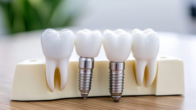
Implant design is critical in the initial healing, integration with the bone, and the long term success of the treatment. A dental implant has threads similar to a screw. The external threads are designed in a way to distribute biting forces in order to minimize the stress to the bone. The implant is made of titanium and the surface of that titanium is treated in a special way to allow better integration with the bone, a very important process called osseointegration, in which the bone mechanically and chemically fuses to the implant surface. The internal design of the implant is also critical to help maintain the strength, integrity, and biological health of the final restoration, ie crown, that will be placed on the implant.
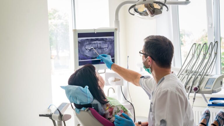
One of the biggest advancements in implant dentistry is the 3D technology available for both planning and execution resulting in a nearly flawless surgical approach. Cone Beam Computed Tomography (CBCT) scans, also generally called a CT scan, allow the dentist and surgeon to develop a customized plan based on each patient’s anatomy. A CT scan is basically a 3-dimensional radiograph. There are times when the patient’s bone is deficient and a bone graft will be necessary. With a CT scan, this can be realized before the patient even makes a decision to move forward. During the planning phase the CT scan allows the dentist to determine all of the procedures and associated costs necessary so that the patient can make an informed decision on whether or not they want to proceed with a dental implant.
After it is determined that there is enough bone for implant placement a surgical plan can be developed. A CT scan and 3D image of the patient’s teeth are used to create a virtual plan of the implant placement. A virtual surgery is completed using special planning software. Once the plan is finalized a surgical guide can be fabricated using a 3D printer. The surgical guide will then be placed in the patient’s mouth at the time of surgery. The surgical guide has a drill sleeve that will guide the direction of the surgery so that the implant is placed at the exact depth and orientation as it was in the 3D plan. This creates a completely custom and individualized approach that results in precise surgical and restorative execution.
Why is this important? Implants are mostly designed to accept forces down the long axis of the implant. The surgical guide allows the precision to place an implant so that the final tooth or crown will be in a position to accept forces down this long axis. Also, the surgical guide helps prevent human error with depth and angulation of placement and will help avoid critical anatomical structures such as the sinus or nerves. We realize the value of this technology for the benefit of the patient. This is why we use surgical guides for every implant placement in our practice. It is important that we take advantage of the wonderful technology we have available and that we continue to improve the clinical outcomes of dental implant treatment.
Any content, resident submissions, guest columns, advertisements, and advertorials are not necessarily endorsed by or represent the views of Best Version Media LLC (BVM) or any municipality, homeowners associations, businesses, or organizations that this publication serves. BVM is not responsible for the reliability, suitability, or timeliness of any content submitted, inclusive of materials generated or composed through artificial intelligence (AI). All content submitted is done so at the sole discretion of the submitting party.